Dermoscopy, confocal microscopy, and optical coherence tomography offer advantages in diagnosing skin cancer, each contributing to non-invasive detection.
As skin cancer rates continue to rise globally, the demand for more effective diagnostic tools has never been greater. Non-invasive imaging technologies such as dermoscopy, confocal microscopy, and optical coherence tomography (OCT) have improved the early detection and management of skin cancers. This article compares these key imaging techniques, highlighting their roles, effectiveness, and how they complement each other in clinical practice.
Dermoscopy: The First Line of Defense
Dermoscopy is often the first step in the skin cancer diagnostic process, especially for melanoma. By magnifying the skin and using polarized light, dermoscopy allows dermatologists to observe pigmentation patterns and vascular structures that are not visible to the naked eye.
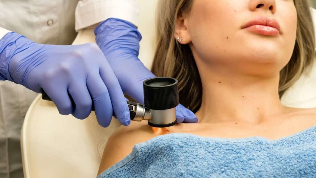
This technique has significantly improved the diagnostic accuracy for melanoma, reducing unnecessary biopsies.
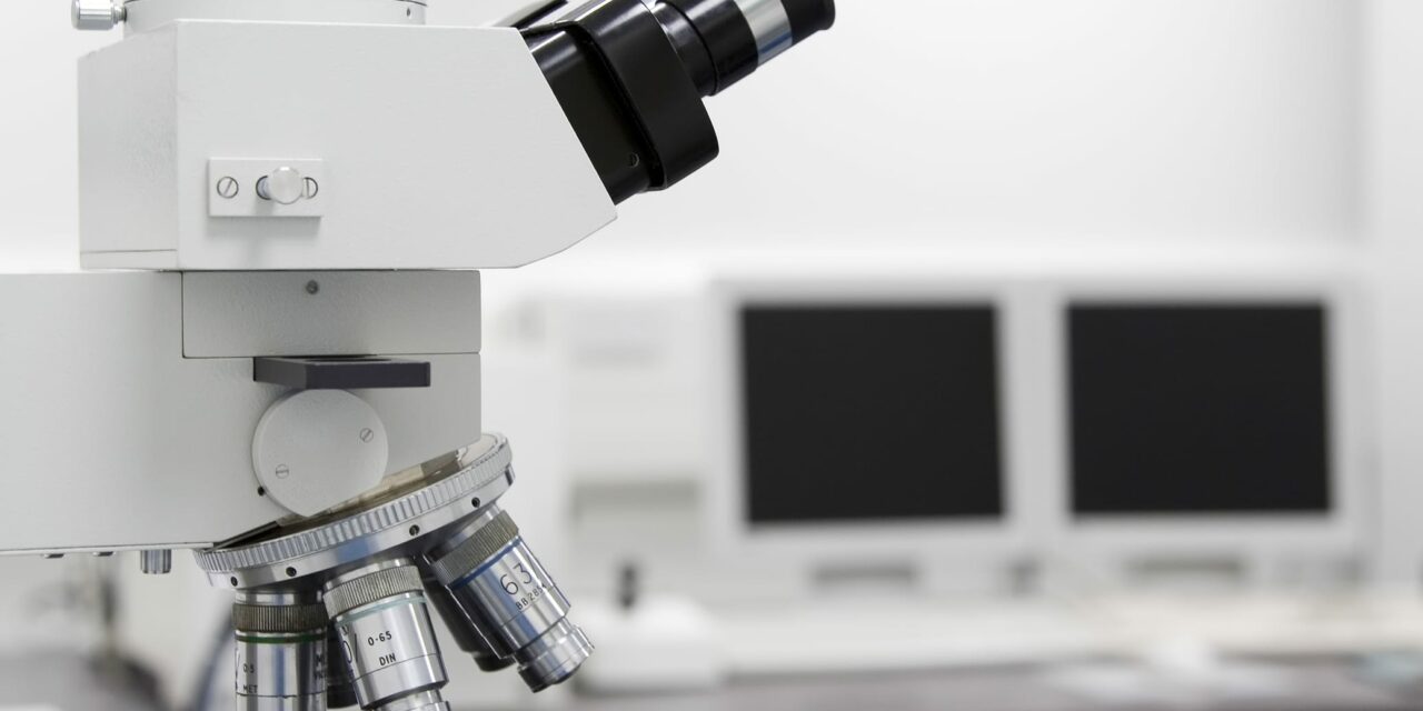
Confocal Microscopy: A Closer Look at Cellular Structures
Confocal microscopy provides a more detailed view at a cellular level, allowing clinicians to examine skin layers non-invasively with nearly histological detail. Particularly effective for diagnosing basal cell carcinoma, confocal microscopy can differentiate between benign and malignant cells without the need for a biopsy, aiding in the decision-making process for treatment strategies.
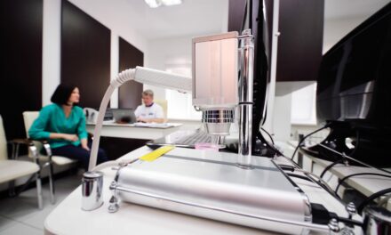
Optical Coherence Tomography: Visualizing Skin Architecture
Optical coherence tomography uses light waves to take cross-sectional images of the skin, providing detailed information about skin architecture. It is particularly useful for identifying non-melanoma skin cancers like basal cell carcinoma. Optical coherence tomography can determine tumor depth and boundaries, which are crucial for treatment planning and monitoring response to therapies.
Comparative Analysis and Clinical Integration
While each of these imaging techniques offers distinct advantages, their integration can provide a comprehensive diagnostic picture. For example, while dermoscopy can suggest a preliminary diagnosis, confocal microscopy and optical coherence tomography can confirm it, reducing the need for invasive procedures. The choice of imaging technique often depends on the type of skin cancer suspected, the lesion’s characteristics, and the patient’s medical history.
The comparative effectiveness of non-invasive imaging techniques has made substantial contributions to the field of dermatology, particularly in skin cancer diagnosis. As these technologies continue to advance, their integration into routine clinical practice is expected to improve, offering more precise diagnostics and better patient outcomes.
Photo 636281 © Waiheng | Dreamstime.com
