Modern dermatoscopic advancements, from enhanced imaging to AI integration, are improving dermatological diagnostics. Explore how these innovations are making a difference in skin cancer detection and management.
Dermatoscopy has significantly evolved, with continuous advancements in technology and techniques enhancing its diagnostic accuracy and utility. These developments are crucial for improving the early detection and management of various skin conditions, particularly skin cancers. This article explores the latest innovations in dermatoscopy and their impact on dermatology practice.
Evolution of Dermatoscopic Devices
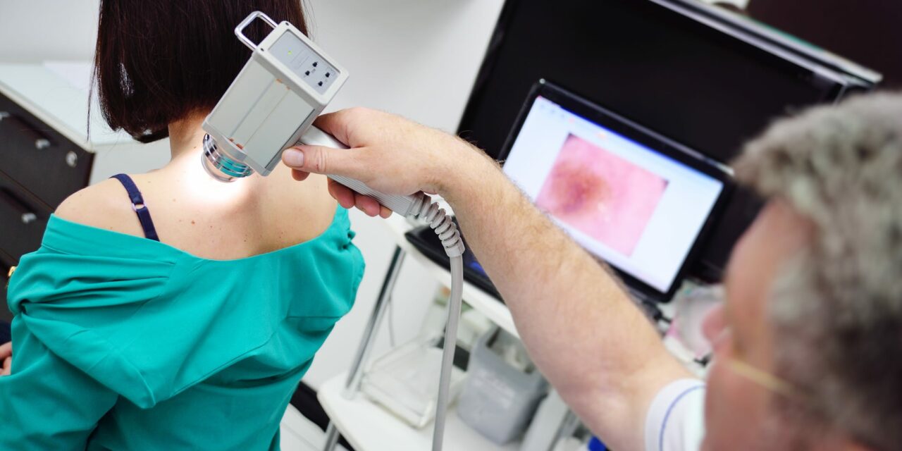
The traditional handheld dermatoscope has undergone substantial modifications, incorporating advanced features that augment its diagnostic capabilities. Modern dermatoscopes now often include polarized light, which reduces surface glare and allows for the visualization of deeper skin structures.
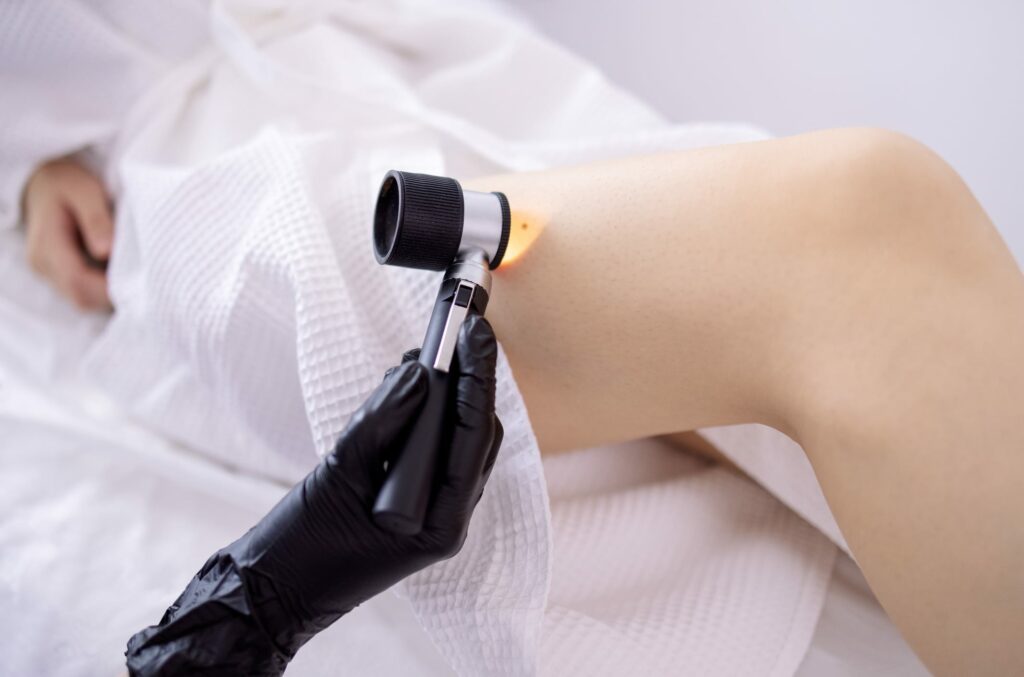
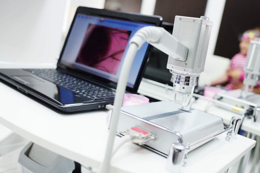
This innovation facilitates better differentiation between benign and malignant lesions. Digital dermatoscopes, which capture and store high-resolution images, enable detailed analysis and long-term monitoring of skin lesions.
Integrating Artificial Intelligence
One of the most transformative advancements in dermatoscopy is the integration of artificial intelligence (AI). AI algorithms, trained on vast datasets of dermatoscopic images, can assist in the identification and classification of skin lesions.
Studies have shown that AI can achieve diagnostic accuracy comparable to experienced dermatologists. For instance, a study published in Nature Medicine demonstrated that a convolutional neural network (CNN) trained on dermatoscopic images achieved a sensitivity of 95% for melanoma detection, outperforming many clinicians^1.
Enhanced Imaging Techniques
High-definition imaging and the use of multispectral analysis have further refined dermatoscopic techniques. Multispectral dermatoscopes analyze the skin at various wavelengths, providing comprehensive information about lesion characteristics. This technique enhances the visualization of pigmentation patterns, vascular structures, and other critical features, aiding in the accurate diagnosis of complex cases.
According to a review in Journal of the American Academy of Dermatology, multispectral imaging significantly improves diagnostic accuracy for both melanocytic and non-melanocytic lesions^2.
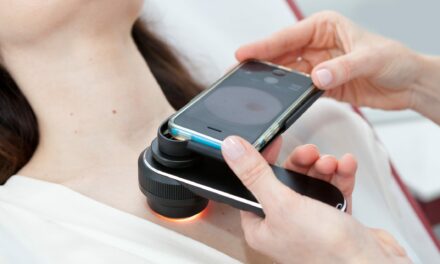
Mobile Dermatoscopy
The advent of mobile dermatoscopy has democratized access to this diagnostic tool. Smartphone attachments and dedicated apps enable dermatologists to perform dermatoscopic examinations in various settings, including remote and underserved areas.

This portability facilitates teledermatology consultations, allowing for expert analysis even when direct patient interaction is not possible. Research published in JAMA Dermatology highlighted that mobile dermatoscopy could bridge the gap in dermatological care in resource-limited regions^3.
Software Innovations and Image Analysis
Advanced software for image analysis and management has also emerged as a crucial component of modern dermatoscopy. These platforms offer features such as lesion segmentation, pattern recognition, and longitudinal tracking. By maintaining a digital record of lesions, dermatologists can monitor changes over time, which is particularly beneficial for patients with multiple atypical moles or those at high risk for skin cancer.
A study in Dermatologic Surgery emphasized the importance of software-assisted dermatoscopy in enhancing diagnostic precision and facilitating patient follow-up^4.
The advancements in dermatoscopic technology and techniques have revolutionized dermatological diagnostics. From AI integration to mobile applications, these innovations have significantly improved the accuracy and accessibility of skin examinations. As technology continues to evolve, dermatologists must stay abreast of these developments to provide the best possible care for their patients.
References
- Tschandl, P., et al. (2020). Human–computer collaboration for skin cancer recognition. Nature Medicine, 26, 1235-1243.
- Malvehy, J., et al. (2007). Dermoscopy report: proposal for standardization. Journal of the American Academy of Dermatology, 57(4), 701-703.
- Wolf, J. A., et al. (2013). Diagnostic inaccuracy of smartphone applications for melanoma detection. JAMA Dermatology, 149(4), 422-426.
- Vestergaard, M. E., et al. (2008). Dermoscopy compared with naked eye examination for the diagnosis of primary melanoma: a meta-analysis of studies performed in a clinical setting. Dermatologic Surgery, 34(5), 587-595.
Photo 103895298 © Evgeniy Kalinovskiy | Dreamstime.com
